


In the golden September, the academic hall shines brightly. The 29th Ophthalmology Academic Conference of the Chinese Medical Association (CCOS 2025) was successfully held at the Hangzhou International Expo Center. At this annual feast in the field of ophthalmology, the vibrant and diverse - the seminar on new technologies for ultra wide angle multimodal fundus imaging was also successfully held at the same time. Yaoshi Medical led the trend with its innovative technology and showcased its flagship ultra wide angle laser scanning ophthalmoscope (SLO) Yetsea 300 in the "Yao · Xinghai" series, becoming the focus of attention for the audience. The "Yao · Xinghai" series laser scanning ophthalmoscope utilizes advanced confocal scanning technology to capture fundus images with unprecedented clarity and breadth, providing doctors with more comprehensive and accurate diagnostic criteria. The flagship Yetsea 300 in this series is a masterpiece after multiple upgrades and innovations, showcasing Yaoshi Medical's outstanding innovation capabilities and solid research and development strength. The International Ophthalmology News specially captured the essence of the seminar on new technologies of ultra wide angle multimodal fundus imaging, and invited Professor Zhao Mingwei from the People's Hospital of Peking University to conduct an exclusive interview to discuss the important clinical value of Yetsea 300.
Driven by innovation, the new technology of ultra wide angle multimodal fundus imaging leads a new era of fundus examination
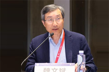

Host: Professor Wei Wenbin from Beijing Tongren Hospital affiliated with Capital Medical University and Professor Zhao Mingwei from Peking University People's Hospital
Unlocking the clinical application of multimodal fundus imaging technology
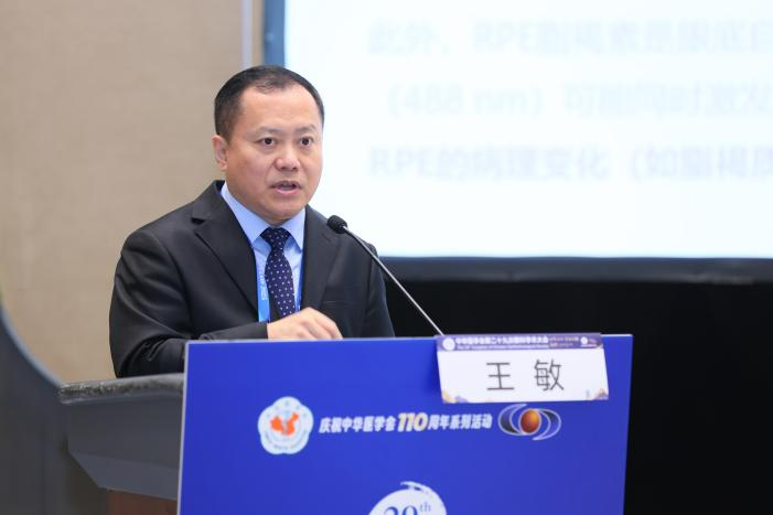
Confocal laser scanning ophthalmoscope has the technical characteristics and advantages of layered imaging, high resolution, and comfortable lighting, and the longer the laser, the better the penetration power. Professor Wang Min from Fudan University Affiliated Eye, Ear, Nose and Throat Hospital introduced that the Yetsea 300 from Yaoshi has achieved synchronized imaging of red, blue, green, and infrared four-color lasers for the first time in China, which can clearly display the fine structure of the retina in layers. One scan can obtain five fold images, making the lesion nowhere to hide. In addition, the Yetsea 300 from Yaoshi uses multi laser confocal technology to capture a retinal range of 168 degrees in a single scan, with images that are close to true color, which helps detect peripheral lesions and displays changes in lesions at different levels, helping clinical doctors understand the condition. The multi-mode integrated design allows for horizontal comparison of lesion manifestations in different imaging modes, while reducing errors in device operation and patient cooperation, and shortening the patient's movement curve. The imaging characteristics of multi laser scanning image synthesis color photos are related to the wavelength of the laser used and the algorithm of image synthesis. There are significant differences between different devices, and in clinical practice, disease diagnosis needs to be combined with other examinations. In summary, ultra wide angle multi wavelength imaging has high application value in the precise diagnosis and staging of retinal diseases such as macular diseases, vascular diseases, congenital diseases, genetic diseases, systemic diseases, trauma, peripheral retinal degeneration, etc.
A Brief Discussion on the Clinical Application Value of Different Wavelength Laser Layered Imaging
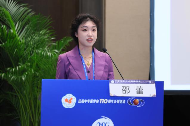
Professor Shao Lei from Beijing Tongren Hospital affiliated with Capital Medical University talked about the development of fundus imaging, pointing out that different wavelength laser layered imaging technology provides ophthalmologists with high-definition images of each layer of the fundus, accurately assisting in disease diagnosis, treatment plan selection, and disease monitoring. CSLO, with its advantages of non mydriasis, strong penetration, and high-definition imaging, has become a powerful tool for early screening of various fundus diseases, winning treatment opportunities for patients. This technology goes deep into choroidal imaging, enhances the diagnostic ability of choroidal diseases, and can dynamically record changes in the fundus, effectively distinguishing between vitreous and retinal lesions. With the advancement of technology and the deepening of clinical applications, cSLO technology will demonstrate more clinical value and lead a new revolution in ophthalmic diagnosis and treatment.
Chairman's comments
Professor Wei Wenbin: The confocal four laser layered scanning technology focuses on the two core areas of scanning area and layered diagnosis, providing important support for the diagnosis of retinal diseases. The Yetsea 300 from Yaoshi can achieve ultra wide angle imaging, with a single scan covering 168 degrees of the retina and presenting approximately 90% of the fundus area. At the same time, with the help of multi wavelength laser scanning, different hierarchical structures of the retina can be clearly displayed, achieving accurate stratification and differential diagnosis. As a comprehensive imaging diagnostic system designed to meet clinical needs, it is expected to play an important role in the screening and accurate diagnosis of retinal diseases.
Professor Zhao Mingwei: In recent years, domestic imaging equipment has made significant progress, represented by brands such as Yaoshi, surpassing some imported products in terms of image clarity, scanning depth, and real-time imaging speed, achieving technological breakthroughs. The Yetsea 300 from Yaoshi has enriched the connotation of imaging diagnosis through innovative multi-mode imaging and multi band layered scanning. In clinical applications, this technology is mainly used in four major scenarios: first, storage and subsequent analysis of imaging data; The second is the diagnosis and differential diagnosis of diseases; Thirdly, guide the selection of treatment methods; The fourth is disease tracking and prognosis judgment. Overall, domestic equipment not only achieves technological breakthroughs, but its multi-mode imaging system also plays an important role in clinical diagnosis and treatment.
In depth interview with Professor Zhao Mingwei, highlighting the significant clinical value of Yetsea 300
Four laser confocal technology is one of the core innovations of Yaoshi Yetsea300. By synchronously exciting lasers of different wavelengths, it achieves synchronous imaging of the shallow deep choroid structure of the retina, which can clearly display different levels of lesions in the fundus, such as key lesions such as microangiomas, neovascularization, choroidal polyps, etc; At the same time, laser dot matrix scanning reduces scattered light interference, which helps to improve the detection of small lesions (such as microangiomas with diameters<50 μ m). This not only helps to clarify the hierarchical location and interrelationships of lesions, but also has the potential to shift the diagnostic model from "single level isolated analysis" to "multi-level correlation verification". The multi-mode integrated design of Yaoxinghai Yetsea300 integrates multiple ultra wide angle imaging modes such as color photography, single laser imaging (B/G/R/IR), spontaneous fluorescence, FFA, and ICGA. This paradigm upgrade is particularly suitable for complex fundus diseases such as PCV and AMD that require multi-level correlation analysis, greatly improving diagnostic accuracy and efficiency. This is not only an expansion of imaging range, but also a revolution in imaging diagnostic thinking.
Yetsea300 from Yaoshi, ushering in a new era of fundus disease screening network
The screening of retinal diseases is extremely important for chronic disease management, and it is urgent to popularize retinal screening at the grassroots level. There are three major bottlenecks in traditional fundus examination equipment: firstly, it relies on professional technicians to operate, making it difficult for primary medical institutions (such as community hospitals and physical examination centers) to allocate full-time personnel, resulting in "equipment available but not usable"; Secondly, the examination process is complex, taking 10-15 minutes per examination, and patients (especially the elderly and children) have low cooperation, making it easy to give up halfway through the examination; The third issue is the high cost of equipment and expensive maintenance expenses. Grassroots institutions are unable to afford or unwilling to use it, resulting in a vicious cycle of "idle equipment - stagnant screening". The Yetsea300 effectively solves these problems through the combination of "fully automatic scanning+ultra wide angle imaging+one click operation" technology. Medical staff can independently complete operations through short-term training, completely breaking away from dependence on professional technicians, making it a reality that "community nurses can also do fundus screening"; The single examination time has been shortened to 3-5 minutes, equivalent to one-third of traditional equipment, significantly improving patient compliance and solving the problem of "difficult to adhere to examinations". In particular, it is of great significance for chronic disease patients (such as diabetes patients) who need regular reexamination, and has truly achieved the goal of chronic disease management of "grassroots screening, screening coverage, coverage can be effective". In the future, if artificial intelligence technology can be deeply integrated for comprehensive analysis and judgment, it will significantly improve the performance of new devices and open up broader market application prospects.
New Vision of Grassroots Ophthalmology: Advanced Equipment Helps Leap in Diagnosis and Treatment Level
Yaoshi Ultra Wide Angle SLO has broken the technological monopoly of imported brands in the field of ultra wide angle fundus imaging, and its cost-effectiveness advantage will promote the improvement of primary hospital fundus disease screening capabilities. From an industry perspective, it marks a leap from "imitation" to "innovation" in China's ophthalmic equipment. When young doctors are exposed to this emerging fundus imaging technology, they need to be careful to avoid "technology for the sake of technology" and clarify three points: first, they need to avoid overly relying on equipment and neglecting clinical examinations; Secondly, it is necessary to understand the imaging differences between laser scanning and optical imaging to avoid improper image interpretation; The third is to establish an imaging clinical correlation, combining ultra wide angle imaging with patient medical history and other examinations (such as OCT) for analysis, to avoid "talking based on pictures". By integrating imaging findings with clinical symptoms and signs, it will help to analyze retinal diseases more comprehensively, thereby enhancing the comprehensive diagnostic and treatment thinking ability of young doctors.
summary
Yaoshi Medical has always adhered to the mission of "bringing brightness and hope to every pair of eyes", constantly exploring and innovating, striving to become a globally trusted leader in eye health solutions with technological innovation as the wing and medical needs as the guide. We hope that the Yetsea 300 from Yaoshi can inject new vitality into Chinese ophthalmic medical care, help Chinese ophthalmologists achieve more efficient diagnosis and treatment processes, and assist patients in obtaining safer and more comfortable diagnosis and treatment experiences.